
 (8621)54893781,64688888-801/810
  info@renai.cn
 Chinese


Breast lump: Early evaluation is essential
If you find a breast lump or other change in your breast, you might worry about breast cancer.That's understandable — but remember that breast lumps are common. Most often they're noncancerous (benign), particularly in younger women. Still, no matter how old you are, it's important to have any breast lump evaluated by a doctor, especially if it's new and feels different from surrounding breast tissue.
How breast tissue normally feels?
Breasts contain tissues of varying consistency. The glandular tissue in the upper, outer part of the breast usually feels slightly rope-like, bumpy or lumpy (nodular). The surrounding fat tissue, often felt in the inner and lower parts of the breast, is soft and less nodular or lumpy than the upper, outer breast.
You might find that breast-related symptoms, such as tenderness or lumpiness, change with your menstrual cycle. Breast tissue also changes as you age, typically becoming more fatty and less dense.
When to consult your doctor?
Being familiar with how your breasts normally feel makes it easier to detect when there's a change in your breasts.
Consult your doctor if:
1. You find a new breast lump.
2. A new breast lump or breast pain doesn't go away after your next period
3. An existing breast lump gets bigger or otherwise changes.
4. You notice skin changes on your breast, such as redness, crusting, dimpling or puckering.
5. You notice changes in your nipple — it turns inward (inversion) or appears flatter, for instance.
6. You notice spontaneous nipple discharge from one breast that's clear, yellow, brown or red.
What to expect during a clinical breast exam?
Evaluation of a breast lump typically begins with a clinical breast exam. During this exam, your doctor will likely:
Ask about symptoms and your risk factors for breast cancer or benign breast conditions.
Examine your breasts, noting their shape and size, while you're standing and while you're lying down.
Examine the skin on your breasts.
Check for nipple problems, such as inversion or discharge.
Feel (palpate) the deeper tissue in your breasts and armpits to detect lumps or areas of thickening.
If your doctor confirms that you have a breast lump or other area of concern, you'll likely need testing.
Procedures to evaluate a breast lump
To further evaluate a breast lump, your doctor might recommend one or more of the following procedures.
Mammogram
A diagnostic mammogram — a specialized breast X-ray — helps your doctor investigate breast lumps and other signs and symptoms, such as tissue thickening, skin dimpling or nipple inversion.
A diagnostic mammogram focuses on one area of your breast, providing views from several angles at higher magnification than does a screening mammogram.
This test helps your doctor pinpoint the location and the size of the abnormality.
A diagnostic mammogram is often done along with an ultrasound of the breast.
Ultrasound
Sound waves create images of the inside of your breast on a monitor. Ultrasound imaging is helpful for determining whether a breast lump is solid or filled with fluid.
MRI
A magnetic field and radio waves create detailed images of the inside of your breast. A breast MRI usually is reserved for when the diagnosis is in question.
When an MRI is used to detect breast cancer, a special dye (contrast agent) must be injected into your veins before the procedure. The dye enhances the appearance of certain tissues in the MRI images, allowing a radiologist to tell which areas are likely to be cancerous.
MRI scans can be challenging to interpret. This can lead to a false-positive result — when the test result is positive but there's no cancer — or the need for additional testing.
Ductogram
Also called a galactogram, this test is sometimes used to find the cause of nipple discharge. A small amount of dye is injected into a duct in the nipple. The dye shows up on an X-ray and can reveal a tumor in the duct.
Breast biopsy
Sometimes removing a tissue sample to examine under a microscope (biopsy) is the only sure way to determine if a breast lump is cancer. The type of biopsy depends on the size and location of the suspicious area.
Breast biopsy options include:
Fine-needle aspiration biopsy
Core needle biopsy
Stereotactic biopsy.
Vacuum-assisted biopsy
Surgical biopsy
All biopsies can cause bruising, bleeding and swelling. A surgical biopsy will likely leave a scar, and depending on how much tissue is removed, may change the shape of your breast.
After a biopsy, the tissue sample is sent to a lab for analysis. Your doctor will let you know when to expect the test results and discuss the results with you when they're available.
Follow-up after breast lump evaluation
If the breast lump isn't cancerous, your doctor might suggest short-term monitoring followed by another clinical breast exam or repeat breast imaging in a few months to reassess the area. Consult your doctor if you notice changes in the lump or develop new areas of concern.
If the diagnosis is in question — the clinical breast exam and the mammogram show areas of suspicion, for example, but the pathology report from the biopsy reveals benign tissue — you'll be referred to a surgeon or other specialist for further consultation.
If the breast lump is cancerous, you'll work with your doctor to create a treatment plan. The stage and type of breast cancer will influence your treatment options. If you're unsure how to proceed, ask your doctor to help you make the best treatment decisions.
- Doctors' Working Schedule from Feb 17 to Feb 28Feb 17, 2025
- Doctors' Working Schedule from October 12 to October 31Oct 12, 2024
- Doctors' Working Schedule from 20th September to 30th SeptemberSep 20, 2024
- Doctors' Working Schedule from 6th September to 18th SeptemberSep 6, 2024
- Doctors' Working Schedule from 15th August to 31st AugustAug 14, 2024
-
-
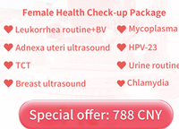
Get Women's Day Special Offer of Health Checkups for Her & H
Time-limited special offer in March.
-

-
Sepcial Offer: Exclusive Health Checkup Packages for Men&Wom
-

-
Time-limited 50% Off! Get Your Annual Health Checkup Here No

 沪公网安备 31010402006742号